The Kennewick Man Case | EventsEstablishing a baseline: Taphonomic studies July 2005Beginning July 6, 2005, a team of scientists conducted a thorough taphonomic analysis the Kennewick Man skeleton at the Burke Museum, University of Washington, Seattle. This ten-day evaluation assessed the evidence of events that have affected the skeleton from its deposition at the time of death to the present. The evidence included the actual bone fragments, the archived image record, and the curation history. A few weeks before the taphonomic studies began in Seattle, high density CT scans were taken of the 9 pieces of the cranium and the fragments of the right hip with the projectile point. Polymer prototyes, made from the scan data, arrived the day the studies began. These pictures show the team at work.
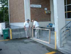
The first morning - UW's Burke Museum loading dock. Constructing the 8 ft x 4 ft sand table used to lay out the skeletal fragments in anatomical order. Shown are Chip Clark, Smithsonian and Stuart Hawkinson, Friends of America's Past.
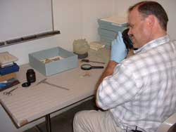
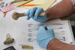
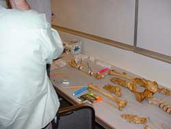
Dr. David Hunt assessed the accuracy of the cranium prototype fragments by compariang them with the actual bone fragments. In this picture, Dr. Hunt photographs a piece of malar (cheek bone) and its prototype. The other fragments of the face are in the blue box to the left of the prototype of the cranial vault (upside down on a cushion). Dr. Hunt then reconstructed the prototype and the cranium to evaluate the accuracy of the prototyping process.
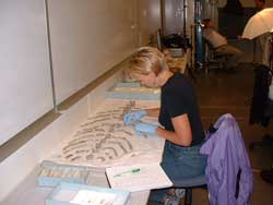
Kari Bruwelheide, Smithsonian osteologist, had the daunting task of fine-tuning the prior identifications and placement of the fragmented ribs. Kari was also able to place most of the many 'unidentified' fragments (blue box with small plastic bags). Creating as complete a layout as possible was a critical first step for the taphonomic analysis.
Dr. Doug Owsley and Dr. Hugh Berryman, fracture expert, spent the first two days evaluating and describing the femur fragments. Alethea Snyder recorded their observations into a spreadsheet. Owsley and Berryman were the first non-Army Corps scientists to assess these fragments after they were returned to the collection in 2001. During his inventory in 1998, Dr. Owsley confirmed rumors that portions of both femurs were missing from the collection. After the Army Corps of Engineers took possession of the collection in 1996, they had stored the bones in an unlocked cabinet. The missing femurs were 'rediscovered' in a Benton County Sheriff Department evidence locker. The taphonomic story for the femurs has a special chapter to be explored.
Hugh Berryman recorded each fracture and its location (anterior and posterior views). He can distinguish old fractures from those that occurred as the body eroded out of the bank. He also assessed the fractures that have resulted from fluctuating temperature and relative humidity since the bones have been stored at the museum.
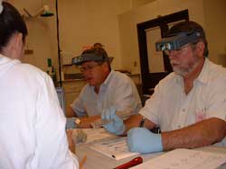
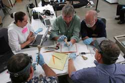
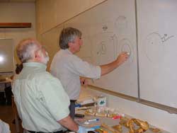
Drs. Stafford, Smith, Owsley, and Berryman discussed their observations as a team, element by element. Alethea Snyder, data analyst, recorded more than two dozen variables on spreadsheets.
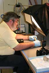
Chip Clark, Smithsonian photographer, took high quality scientific photos. Many of these photos will be published with the final scientific reports and will be an invaluable record for future scientists.
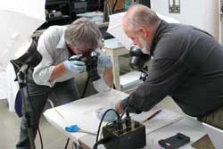
Drs. Stafford and Smith photographed the locations of calcium carbonate concretions on fragment surfaces. When possible they also photographed the crystals inside fractured shafts. The formation of these crystals reveals important clues about the position of the bone in the ground.
The team used a disarticulated skeleton with unbroken bones to help visualize the fracture patterns on the Kennewick fragments. Using pipe cleaners, colored post-it notes, and plastic stirring sticks, they recreated the angles of force that caused the fractures.
Dr. Stafford, geochemist, evaluated the chemical deposition patterns and Dr. Smith, a specialist on the effects of water on bone discussed the implications of their observations. The goal was to gain insights about the original position of the body in the ground and the order in which the bones eroded from the bank.
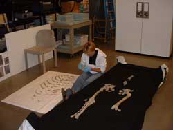
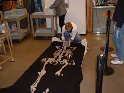
After finalizing their identifications, Kari began to lay out the fragments in anatomical order on the sand table. Special black fabric covered bout 400 pounds of sand. Sand supports each fragment in anatomical position.
Kari used one hand to hold a cervical vertebra and the other to adjust the sand under the fabric to reproduce the natural curve of the neck.
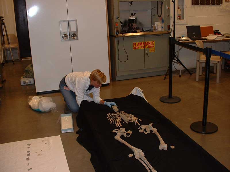
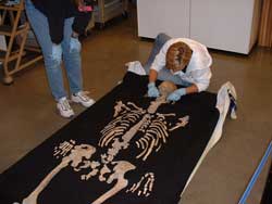
Kari had spent more than a day on the floor, carefully transferring the fragments to the sand table. She arranged and rearranged the sand to place the cranial vault so it would rest securely.
Kari took extra care positioning the face fragments in relation to the cranial vault. The final step was to place the mandible (lower jaw). Although sand supports each fragment, the face was in pieces, so this was an extremely difficult task.
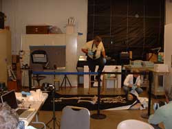
As the scientists' request to video tape in the lab was denied, Chip took photos documenting the entire layout process. After assessing the requirements for the final picture, a very tall ladder was needed to get high enough for the wide angle photos. The final layout photograph will be released in the months to come.
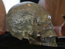
On Sunday, July 10, 2005, the scientists spent the afternoon meeting with the media to talk about their first week of studies and to show them the reconstructed prototypes of the Kennewick Man's cranium and hip. The reconstructed polymer prototype of the cranium was the first use of this technology on human bone. In the months to come, casts will be made so many scientists and students can benefit from the nine-year effort to preserve these precious remains. The projectile point prototype has been digitally 'freed' so that lithic experts can actually hold, rotate, and discuss the type of point still embedded in the bone. The reconstructed prototype of the hip will resolve the controversy about the direction from which the point entered the bone. Photo credits go to the various team members. We offer special thanks to Dr. Nathan Myhrvold, whose generous gift made it possible for the scientists to pursue, with great success, the prototyping process. We also thank Art Andersen, Virtual Surfaces Inc., and Brian Wilcox, Point Data Marketing, Inc., and Mobile Scanning Laboratory, Inc., for their invaluable skills to deliver the prototypes on an extremely a tight schedule. Return to Events |
||